Squamous Cell Skin Cancer
Learn about squamous cell carcinoma, its causes, symptoms, and treatment options. Discover the importance of early detection and effective treatments for healthy skin.
What is Squamous Cell Carcinoma?
Squamous cell carcinoma is the second most common type of skin cancer. Over 1 million new cases of cutaneous squamous cell carcinoma are diagnosed annually in the United States.
Squamous cell carcinoma is an abnormal growth of the squamous cells of your skin. It can be a dangerous type of skin cancer. It can spread (metastasize) to your lymph nodes or distant sites, but thankfully, this is rare, occurring in 2% to 5% of these cancers. Most squamous cell carcinomas have a cure rate greater than 98% if treated early. If these skin cancers are not treated early or correctly, they can spread (metastasize).
In the Southern half of the United States, there are more skin cancer deaths from squamous cell carcinoma than melanoma. This is due to the large amount of sun exposure in the Southern United States. Patients who have organ transplants or Chronic Lymphocytic Leukemia are more likely to develop squamous cell skin cancers and should be seen by a Board-Certified Dermatologist regularly.
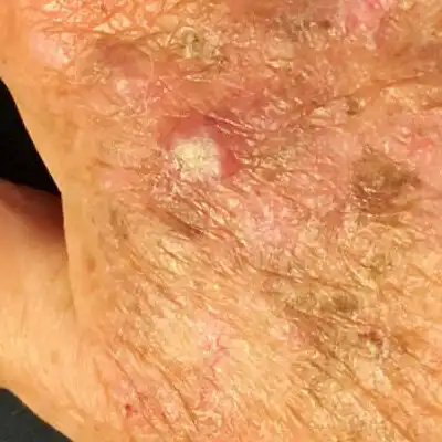
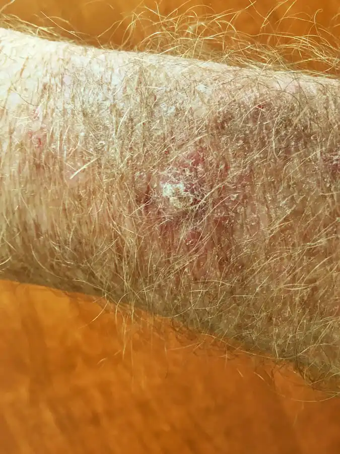

What Does Squamous Cell Carcinoma Look Like?
Squamous cell carcinomas most often occur in areas with frequent sun exposure, such as the face, scalp, neck, ears, backs of the hands, forearms, and shins. However, they can also occur in many other areas of the skin. Squamous cell carcinoma often looks like a rough, scaly spot, scab, or sore that will not heal. It tends to bleed more easily than normal skin.
Scabs and scratches typically heal within 4 weeks. Skin cancer does not heal because the cells are abnormal. If you have a non-healing scab or sore that has been present for more than 4 weeks, you should call your dermatologist to examine it.
It is imperative to note that while these characteristics are some of the most common signs of squamous cell carcinoma, there is no substitute for your skin concern being examined in person by a Board-Certified Dermatologist. This is the gold standard for addressing any of your skin concerns. Your Board-Certified Dermatologist will perform a comprehensive examination and is the expert in diagnosing and treating skin cancers.
What Causes Squamous Cell Carcinoma?
- Cumulative sun exposure over your entire life.
- Tanning beds or sunlamps
How is Squamous Cell Carcinoma Diagnosed?
Squamous cell carcinoma is diagnosed with a skin biopsy. A skin biopsy is a minor procedure in which your dermatologist numbs the area and then takes a sample of your lesion. This sample is examined microscopically for the presence of skin cancer cells. If skin cancer cells are seen, further treatment is required.
What is the Best Treatment for Squamous Cell Carcinoma?
The best treatment for squamous cell carcinoma depends on several factors: the type of squamous cell carcinoma, the location of the skin cancer, and your health and medical conditions. Your dermatologist can assess your skin cancer and develop an individualized treatment plan to meet your goals and specific needs.
When appropriate, Mohs surgery offers the highest cure rate among all treatments. 99% of all squamous cell carcinomas can be cured with Mohs Surgery. This technique preserves your normal skin, removing only the cancer, minimizing scarring, and maximizing your cosmetic outcome. Mohs surgery is the most advanced treatment for squamous cell carcinoma.
When is it appropriate to treat a squamous cell carcinoma with Mohs Surgery?
- If it is located on your face, neck, scalp, hands, feet, or shins (any size or type)
- If it is an aggressive subtype of squamous cell carcinoma
- If it is greater than 2 cm (0.78 inches), located anywhere on your body
Did You Know:
- You can use your smartphone to take photos of your skin to document your moles, both how they look and their location. Then, if in the future you are concerned about a spot that has changed, you can refer back to those high-quality photos.
Mohs Surgery & Mohs Procedure St. Louis
Mohs Micrographic Surgery offers the highest surgical cure rate among all treatments for skin cancer. This technique preserves your normal skin, focusing on removing skin cancer, minimizing scarring, and maximizing your cosmetic outcome. Mohs Micrographic surgery is the most advanced treatment for skin cancer.
Mohs Micrographic Surgery is the only surgical technique that examines 100% of the margins (edges) of the specimen (skin cancer) removed. This means that 100% of the specimen’s side and bottom edges are examined microscopically to confirm that there are no cancer cells at the edges. This provides patients with the highest cure rates, the fewest scars, and the best cosmetic outcomes for their skin cancer.
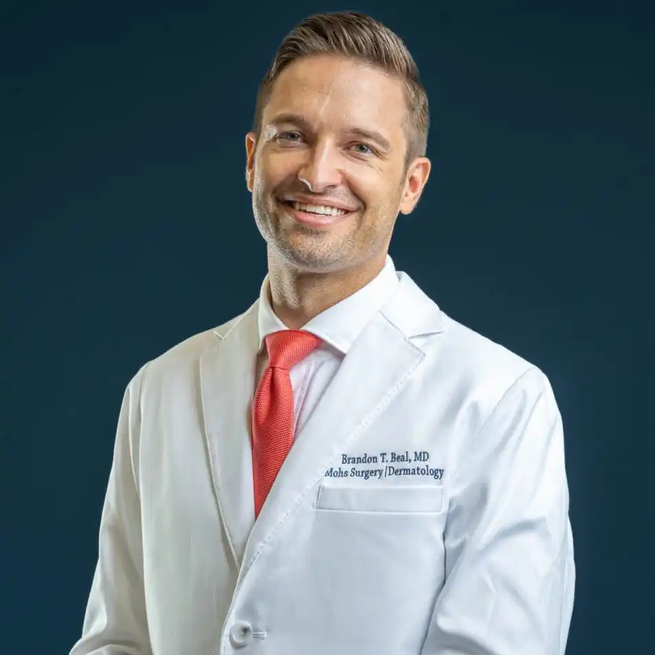
Brandon T. Beal, MD, is an expert in diagnosing and treating skin cancer. He is a Double Board-Certified Mohs Surgeon and Dermatologist, a fellowship-trained Mohs Micrographic Surgeon, a dermatologic oncologist (cancer doctor of the skin), and a plastic facial reconstructive surgeon.
Dr. Beal completed his dermatology residency at the Cleveland Clinic Dermatology & Plastic Surgery Institute and his fellowship in Mohs Micrographic Surgery, dermatologic oncology, and facial plastic and reconstructive surgery at Zitelli & Brodland, PC. Dr. Beal trained at the Cleveland Clinic’s Melanoma program, a multidisciplinary team of dermatopathologists, pathologists, dermatologists, and surgical, medical, and radiation oncologists.
Dr. Beal provides each patient with comprehensive counseling on the diagnosis and treatment of skin cancer, a thorough head-to-toe skin examination, and an individualized treatment plan based on evidence-based medicine. Dr. Beal is an expert in Mohs Micrographic Surgery (Mohs Surgery), which is the treatment that provides the highest cure rates, greater than 99% for most skin cancers. He follows the National Comprehensive Cancer Network Guidelines and the American Joint Committee on Cancer staging guidelines.
GET IN TOUCH