Melanoma Skin Cancer
Learn about melanoma, its causes, symptoms, and treatment options. Discover the importance of early detection and effective treatments for better skin health.
What is Melanoma?
Melanoma is the third most common type of skin cancer. There are over 200,000 new cases of melanoma diagnosed annually. The abnormal growth of melanocytes causes melanoma, the skin cells that produce moles and pigment. Fifty percent of melanomas originate from moles, and the remaining 50% are found in normal skin.
Melanoma is the deadliest skin cancer. 75% of skin cancer-related deaths are due to melanoma. This is because invasive melanomas can spread (metastasize). The ability of a melanoma to spread is directly related to its depth or thickness. Deeper melanomas (> 1mm thick) are more likely to spread beyond the skin to other organs. This is why early detection of melanoma is so essential. Board-certified dermatologists are the experts in the diagnosis and treatment of melanoma. Treatment success is directly related to the depth of the melanoma. Thankfully, when melanoma is detected early, the 5-year survival rate in the United States is 98%.
Melanoma Symptoms (What Does Melanoma Look Like?):
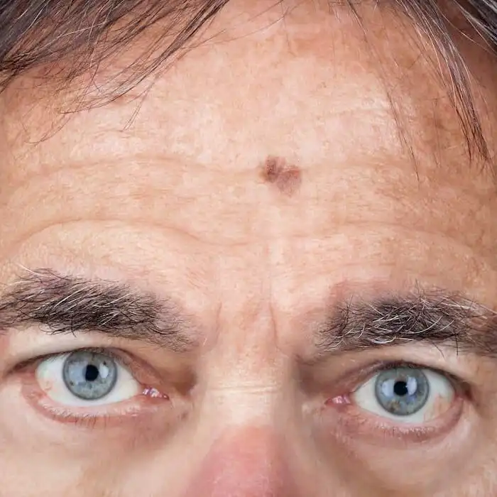
Melanoma often looks like a brown spot or mole that has changed:
- Grown larger (wider or thicker)
- Ulcerated/bleeds more easily than your other moles
- Changed color (ex., became darker)
Melanomas can occur anywhere on the skin. In women, melanoma most commonly occurs on the legs and arms. In men, melanomas are more common on the trunk. As we age, melanomas become more common on the face and in sun-exposed areas of the body.
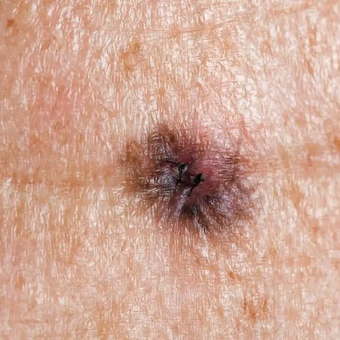
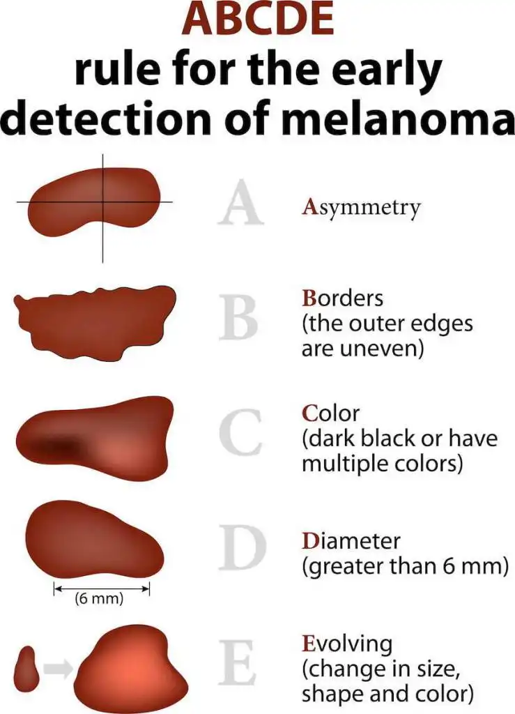
The ABCDEs of melanoma are crucial in helping you identify melanoma early.
A = Asymmetry: is the mole symmetric, one side matches the other
B = Border irregularity: are the borders regular/smooth (reassuring) or jagged and irregular
C = Color: Does the mole or brown spot have the same color (reassuring) or many different colors (brown, blue, black, white or red)
D = Diameter: is the mole the size of a pencil eraser or smaller (<6mm)
E = Evolution: this is the most important. Is the mole changing? If your mole has changed, you should schedule an appointment with a dermatologist.
The Most Common Areas People Forget to Check for Melanoma:
When examining your skin, be sure to check:
- Scalp – it can be challenging if you have a lot of hair, so consider asking a friend or your hairdresser for help.
- Bottoms of your feet and in between your toes
- Back and the back of your legs – Tip: use a full-length mirror and a hand mirror
How to Take High-Quality Photos with Your Phone:
- Ensure you are in a well-lit room with ample lighting. Natural light is the best.
- Take your time and make sure the photos are in focus. When you zoom in, the images should still be clear and not blurry. If the picture is blurry when you zoom in, the camera may not have focused on the correct area.
- Then, take photos of the entire area, such as the left upper back, left lower back, right upper back, right lower back, left upper arm, left lower arm, and so on.
- Next, take a close-up photo of the area of concern. We recommend taking close-up pictures with your camera, 1 foot away from the lesion. A common mistake is to have the camera too close to the spot of concern.
- After taking these photos, review them and zoom in to ensure they are not blurry.
Risk Factors for Melanoma
- Sun exposure without sun protection
- Having two or more severe/blistering sunburns
- Tanning beds or sunlamps
- Genetics – if you have a family member with melanoma, you are at a higher risk of developing melanoma.
- Having greater than 50 moles
Melanoma Facts:
- Melanoma is the most common cancer in people aged 25-29
- Greater than 50% of melanomas do not come from pre-existing moles. In other words, 50% of melanomas originate from seemingly normal skin.
- The FDA has categorized indoor tanning as a human carcinogen, meaning tanning beds cause cancer in humans.
- You can use your smartphone to take photos of your skin to document your moles, including their appearance and location. Then, if you are concerned about a spot in the future, you can refer back to those high-quality photos.
How is Melanoma Diagnosed?
Melanoma is diagnosed with a skin biopsy. A skin biopsy is a minor procedure in which your dermatologist numbs the area and then takes a sample of your lesion. This sample is examined microscopically for skin cancer cells. If skin cancer cells are seen, further treatment is required.
What is the Best Treatment for Melanoma?
The best treatment for melanoma depends on several factors, including:
- Depth or thickness of the melanoma
- Type of melanoma
- Location of the melanoma
- Your health & medical conditions
Melanoma can be a scary diagnosis, so you need to discuss this diagnosis with an expert:

Brandon T. Beal, MD, is an expert in diagnosing and treating melanoma. He is a Double Board-Certified Mohs Micrographic Surgeon and Dermatologist and a Fellowship Trained Plastic & Reconstructive Surgeon.
Dr. Beal provides each patient with comprehensive counseling on the diagnosis and treatment of melanoma, a thorough head-to-toe skin examination, and an individualized treatment plan based on evidence-based medicine. Dr. Beal is an expert in Micrographic Surgery (Mohs Surgery) for melanoma, which offers the highest cure rates and lowest local recurrence rates for head and neck melanoma. He follows the National Comprehensive Cancer Network Guidelines and the American Joint Committee on Cancer staging guidelines.
Dr. Beal completed a fellowship in Mohs Micrographic Surgery, Dermatologic Oncology, and Facial Reconstructive Surgery, with a focus on comprehensive diagnosis and treatment of melanoma. He also received training at the Cleveland Clinic’s Melanoma program, a multidisciplinary team of dermatopathologists, pathologists, dermatologists, and surgical, medical, and radiation oncologists.
More About Mohs Micrographic Surgery:
Mohs Micrographic Surgery offers the highest surgical cure rate among all treatments for head and neck melanoma. This technique preserves normal skin while focusing on removing the melanoma, thereby minimizing scarring and maximizing cosmetic outcomes. Mohs Micrographic surgery is the most advanced treatment for head and neck melanoma.
Mohs Micrographic surgery examines 100% of the margins (edges) of the specimen removed. This means that 100% of the specimen’s side and bottom edges are examined microscopically to ensure there are no melanoma cells at the edges. Mohs Micrographic surgery provides patients with the highest cure rates, the fewest minor scars, and the best cosmetic outcomes.
Your melanoma treatment, including Mohs Micrographic Surgery, immunohistochemical stains, and reconstruction, is performed and completed on the same day at our office. The procedure is done under local anesthesia, which increases our patients’ safety and comfort.
GET IN TOUCH