Mohs Surgery & Mohs Procedure
Mohs Surgery is a state-of-the-art treatment for skin cancer, offering cure rates greater than 99% for most skin cancers.


What Is Mohs Micrographic Surgery?
Mohs Micrographic Surgery (often called Mohs Surgery) is the most effective surgical technique for treating skin cancer.
It offers the highest cure rate — up to 99% — while preserving as much healthy skin as possible for the best cosmetic outcome.
- The goal of Mohs Surgery is simple:
- Remove 100% of the cancer
- Preserve as much normal tissue as possible
During Mohs, 100% of the margins (edges) of the removed tissue are examined under a microscope. This ensures no cancer cells remain — giving you the highest possible cure rate and smallest possible scar.
Who Performs Mohs Surgery?
Mohs Surgery is performed by Board-Certified Dermatologic Surgeons who have completed additional fellowship training in Mohs Micrographic Surgery and Facial Reconstruction.
A Mohs Surgeon serves as:
- Repairing the wound to achieve the best cosmetic result
- Removing the visible skin cancer
- Examining the tissue under a microscope for remaining “roots” of cancer
Why Choose Mohs Surgery?
Mohs Surgery is considered the gold standard for treating many types of skin cancer, especially those on the face, ears, neck, hands, and other visible or delicate areas.
Advantages of Mohs Surgery:
- Cure rates up to 99%
- Best cosmetic results with minimal scarring
- Same-day treatment and reconstruction
- Performed under local anesthesia — safer and more cost-effective than hospital surgery
- Preserves healthy tissue by removing only what’s necessary
Mohs Surgery vs. Traditional Excision
| Mohs Surgery | Traditional Surgery |
|---|---|
| Examines 100% of tumor edges under the microscope | Examines only a few slices (“bread loaf” method) |
| Up to 99% cure rate | Often less than 90% for facial cancers |
| Immediate results | Results can take up to a week |
| Smaller scars and better cosmetic outcomes | Larger margins and higher recurrence risk |
| Local anesthesia and same-day reconstruction | Usually performed in the OR under general anesthesia |
Patient Experience & Safety Benefits
At St. Louis Dermatology & Cosmetic Surgery, Mohs procedures are performed safely and comfortably in our office — not a hospital.
Here’s why that matters:
- Local anesthesia – safer and easier than general anesthesia
- More affordable – office-based surgery can be up to 25x less expensive than hospital procedures
- Convenient – your surgery and reconstruction are completed in a single visit
- No fasting required – eat breakfast and take your regular medications
- Safe for patients on blood thinners – bleeding is easily managed, while reducing the risk of stroke or heart attack
- Most patients drive themselves home unless surgery is near the eye
This patient-centered approach means you receive precise, safe, and cost-effective care without the stress of hospital surgery.
How Is Mohs Surgery Performed?
Mohs Micrographic Surgery is performed in carefully planned stages, all completed in a single visit under local anesthesia. This process allows your surgeon to remove the cancer precisely while preserving as much healthy tissue as possible.
- Step 1: Local Anesthesia and Tumor Removal
- Step 2: Tissue Processing
- Step 3: Microscopic Examination
- Step 4: Additional Stages (If Needed)
- Step 5: Reconstruction
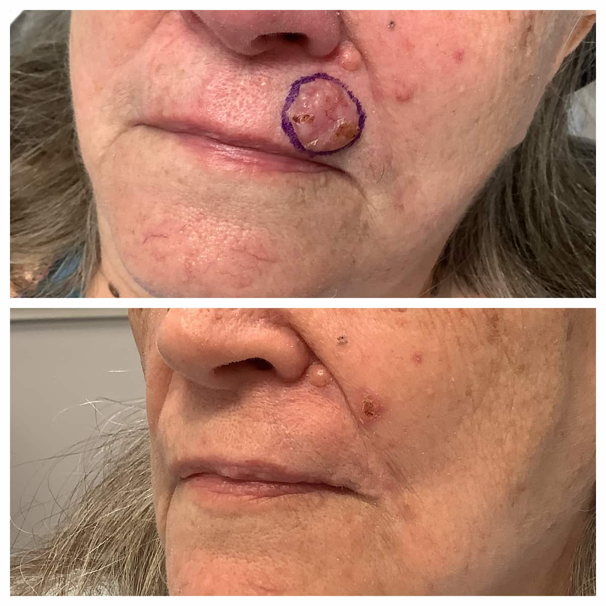
Stage 1: Skin Cancer Removal
Your Mohs Surgeon will begin by reviewing your diagnosis and discussing the procedure in detail. After carefully examining the area, the skin around your cancer is numbed with local anesthesia to ensure your comfort.
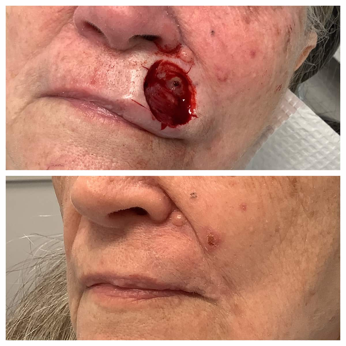
The visible portion of the skin cancer is then removed with a scalpel along with a thin layer of surrounding tissue.
This tissue is inked with colored dyes and mapped like a clock dial (12 o’clock, 3 o’clock, 6 o’clock, and 9 o’clock).
This precise mapping allows your surgeon to know the exact location of any remaining cancer cells seen later under the microscope.
Stage 2: Tissue Processing in Our CLIA-Certified Laboratory
Your specimen is processed immediately in our Centers for Medicare and Medicaid Services (CMS) CLIA-certified laboratory, located on-site. Our specialized technician prepares your tissue for microscopic evaluation — a process that typically takes 45 minutes to 2 hours, depending on complexity. By contrast, traditional surgery may take up to a full week to receive pathology results.
Stage 3: Microscopic Examination
Using a high-powered microscope, your Mohs Surgeon examines 100% of the tissue margins — including both the sides and underside of the specimen. Every edge is checked to ensure there are no remaining cancer cells at the margin.
Because the specimen was precisely mapped, if microscopic tumor “roots” are found, your surgeon can identify their exact location. This allows the removal of only the affected area while preserving normal, healthy skin.
Stage 4: Additional Stages (If Needed)
If microscopic skin cancer remains, only those specific areas are re-excised, and the tissue is reprocessed and re-examined.
This step-by-step process continues until no cancer cells remain, ensuring complete removal while minimizing unnecessary tissue loss.
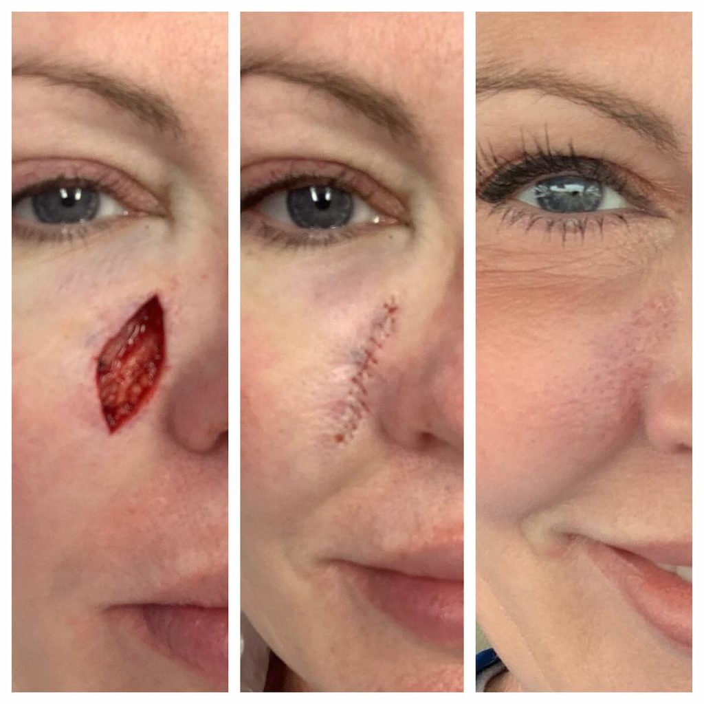
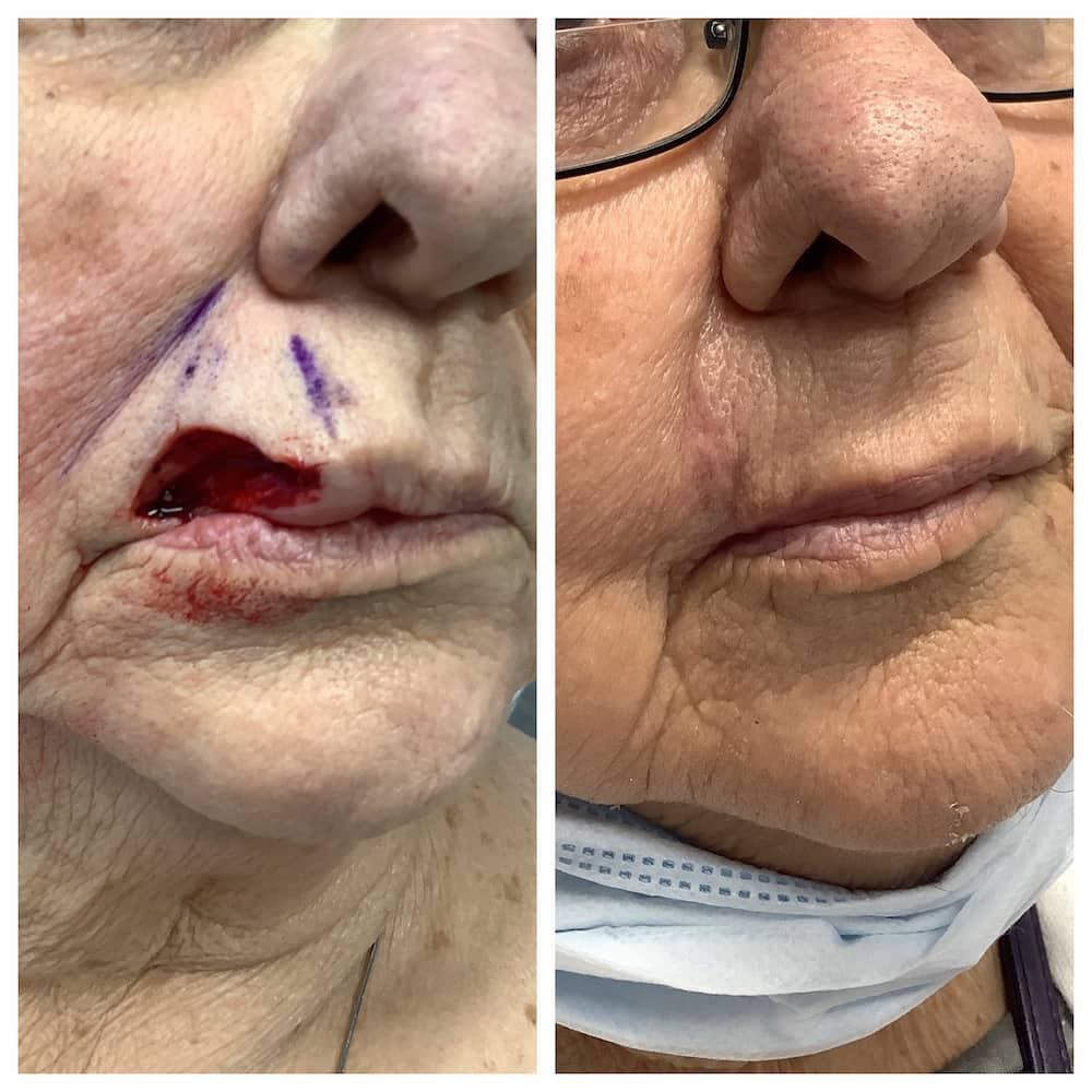
Stage 5: Reconstruction
Once the cancer is fully removed, your Mohs Surgeon will assess your wound and facial appearance to determine the best reconstructive approach.
Restoring Form & Function:
At St. Louis Dermatology & Cosmetic Surgery, our goal is to restore natural form and function — maintaining skin contour, color, and texture for the most aesthetic outcome possible. Mohs Surgeons are highly trained in facial reconstructive surgery and perform more complex facial repairs than any other surgical specialty.
Approved for the Treatment of Rare Skin Cancers
Mohs Micrographic Surgery is approved for the treatment of rare or aggressive skin cancers, including:
- Melanoma and Melanoma in Situ
- Adenocystic carcinoma
- Adnexal carcinoma
- Apocrine/Eccrine carcinoma
- Angiosarcoma
- Atypical fibroxanthoma
- Dermatofibrosarcoma protuberans (DFSP)
- Extramammary Paget’s Disease (EMPD)
- Leiomyosarcoma
- Malignant fibrous histiocytoma / Undifferentiated pleomorphic sarcoma
- Merkel cell carcinoma
- Microcystic adnexal carcinoma (sclerosing sweat duct carcinoma)
- Mucinous carcinoma
- Sebaceous carcinoma
- Other rare cutaneous malignancies
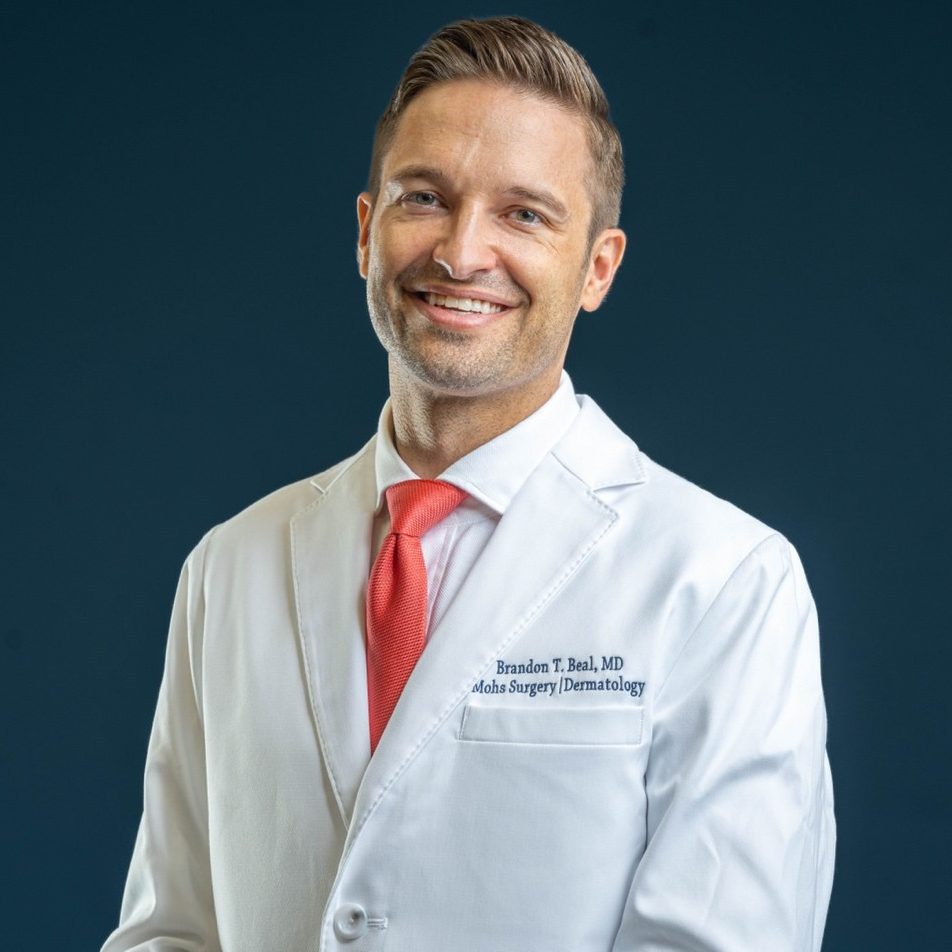
Dr. Brandon T. Beal is a Double Board-Certified Mohs Micrographic Surgeon and Dermatologist, specializing in the diagnosis, treatment, and reconstruction of skin cancer. He completed his dermatology residency at the Cleveland Clinic Dermatology & Plastic Surgery Institute and a fellowship in Mohs Micrographic Surgery, Dermatologic Oncology, and Facial Plastic & Reconstructive Surgery at Zitelli & Brodland, PC — one of the nation’s leading programs.
Dr. Beal’s training also includes work with the Cleveland Clinic Melanoma Program, a multidisciplinary team of dermatologists, oncologists, and pathologists dedicated to advanced skin cancer care.
At St. Louis Dermatology & Cosmetic Surgery, Dr. Beal provides each patient with:
- Comprehensive evaluation and diagnosis
- Individualized, evidence-based treatment planning
- Expert Mohs Surgery and facial reconstruction
- Compassionate, patient-centered care
Dr. Beal follows the National Comprehensive Cancer Network (NCCN) and American Joint Committee on Cancer (AJCC) guidelines to ensure every patient receives care that is precise, effective, and guided by the highest medical standards.
GET IN TOUCH