Home / Medical Dermatology / Mohs Surgery
Mohs Surgery St. Louis
Mohs micrographic surgery removes skin cancer stage by stage, with 100% of the margins examined under the microscope during your visit — cure rates up to 99% for many primary skin cancers, while preserving as much healthy skin as possible.
Double board-certified
Dermatology and Mohs micrographic surgery
Residency & Fellowship
Cleveland Clinic · Zitelli & Brodland
On-site CLIA-certified lab
Margins read during your visit, not next week
Physician-owned
Independent practice — never corporate
What is Mohs Micrographic Surgery?
Mohs micrographic surgery (often simply called Mohs surgery) is a precise surgical technique for treating skin cancer. Its goal is twofold: remove all of the cancer, and preserve as much normal tissue as possible.
What sets Mohs apart is that 100% of the margins — the edges and underside of the removed tissue — are examined under a microscope during your visit. Tissue is removed in stages, with each stage checked before the next, until no cancer cells are seen at the margins. This is why Mohs offers cure rates up to 99% for many primary skin cancers while removing the least tissue necessary.
Who Performs Mohs Surgery?
Mohs surgery is performed by board-certified dermatologic surgeons with additional fellowship training in Mohs micrographic surgery and facial reconstruction. During the procedure, your Mohs surgeon serves three roles: the surgeon removing the visible skin cancer, the pathologist examining the tissue under the microscope for remaining “roots” of cancer, and the reconstructive surgeon repairing the wound.
When is Mohs Recommended?
Mohs is the reference-standard technique for many skin cancers on the face, ears, nose, lips, eyelids, neck, and hands — areas where sparing healthy tissue matters most — as well as for recurrent tumors and aggressive subtypes. Your dermatologist will recommend whether Mohs or another treatment is the right approach for your specific cancer, guided by national treatment criteria.
How is Mohs Surgery Performed?
The entire procedure — removal, margin reading, and reconstruction — is completed in a single visit under local anesthesia, in carefully planned stages.
1
Anesthesia & Tumor Removal
Your surgeon reviews your diagnosis and walks you through the procedure. The skin around the cancer is numbed with local anesthesia, and the visible cancer is removed with a thin layer of surrounding tissue. The specimen is inked with colored dyes and mapped like a clock dial, so any remaining cancer cells can be located precisely later.
2
On-Site Lab Processing
Your tissue is processed immediately in our CMS CLIA-certified laboratory, located right in our office. A specialized technician prepares the slides — typically 45 minutes to 2 hours depending on complexity — while you wait comfortably. With traditional surgery, pathology results can take up to a week.
3
Microscopic Margin Exam
Your surgeon examines 100% of the tissue margins — every edge and the underside — under a high-powered microscope. Because the specimen was precisely mapped, any microscopic “roots” of cancer can be pinpointed, and only those exact areas are re-excised and re-examined. Stages repeat until no cancer cells are seen at the margins.
4
Same-Day Reconstruction
Once the cancer is fully removed, your surgeon assesses the wound and your facial appearance to choose the reconstructive approach — with the goal of restoring natural contour, color, and texture. Mohs surgeons are trained extensively in facial reconstructive surgery, and your repair is completed the same day.
| Mohs Micrographic Surgery | Traditional Excision | |
|---|---|---|
| Margin examination | 100% of the tissue edges and underside, under the microscope | A few representative slices (“bread-loaf” sectioning) |
| Reported cure rates | Up to 99% for many primary skin cancers | Lower — particularly for facial skin cancers |
| Pathology results | Read on-site during your visit | Can take up to a week |
| Tissue removed | Only what is necessary — healthy skin is preserved | Larger, pre-determined margins |
| Setting & anesthesia | Local anesthesia in our office, with same-day reconstruction | Often an operating room, sometimes under general anesthesia |
Individualized careTreatment recommendations are individualized, and results vary. A consultation is required to determine whether Mohs surgery is the right approach for your skin cancer.
In Our Office, Not a Hospital
Mohs procedures are performed safely and comfortably in our office. For most patients, that matters in practical ways:
- Local anesthesia — no general anesthesia and the risks that come with it
- Office-based surgery can cost up to 25 times less than the same procedure in a hospital
- Surgery and reconstruction are completed in a single visit
- No fasting required — eat breakfast and take your regular medications
- Patients on blood thinners typically continue them, avoiding the risks that come with stopping
- Most patients drive themselves home, unless surgery is near the eye
Approved for the Treatment of Rare Skin Cancers
Beyond basal and squamous cell carcinoma, Mohs micrographic surgery is approved for the treatment of rare or aggressive skin cancers, including:
- Melanoma and melanoma in situ
- Adenocystic carcinoma
- Adnexal carcinoma
- Apocrine/eccrine carcinoma
- Angiosarcoma
- Atypical fibroxanthoma
- Dermatofibrosarcoma protuberans (DFSP)
- Extramammary Paget’s disease (EMPD)
- Leiomyosarcoma
- Malignant fibrous histiocytoma / undifferentiated pleomorphic sarcoma
- Merkel cell carcinoma
- Microcystic adnexal carcinoma (sclerosing sweat duct carcinoma)
- Mucinous carcinoma
- Sebaceous carcinoma
- Other rare cutaneous malignancies
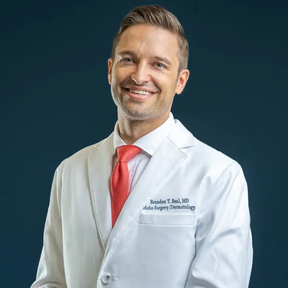
Your Mohs Surgeon: Dr. Brandon T. Beal, MD
Dr. Brandon T. Beal is a double board-certified Mohs micrographic surgeon and dermatologist, specializing in the diagnosis, treatment, and reconstruction of skin cancer. He completed his dermatology residency at the Cleveland Clinic Dermatology & Plastic Surgery Institute and a fellowship in Mohs micrographic surgery, dermatologic oncology, and facial plastic & reconstructive surgery at Zitelli & Brodland, PC — one of the nation’s leading programs.
His training also includes work with the Cleveland Clinic Melanoma Program, a multidisciplinary team of dermatologists, oncologists, and pathologists dedicated to advanced skin cancer care. Dr. Beal follows National Comprehensive Cancer Network (NCCN) and American Joint Committee on Cancer (AJCC) guidelines, so every treatment recommendation is grounded in current national standards.
Every patient receives a comprehensive evaluation, an individualized, evidence-based treatment plan, and compassionate, patient-centered care — from diagnosis through reconstruction and follow-up.
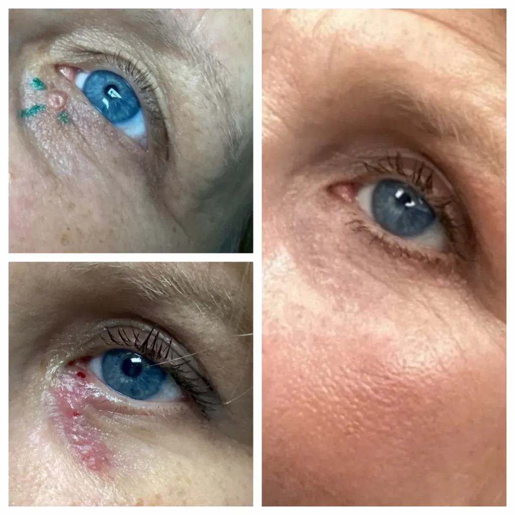
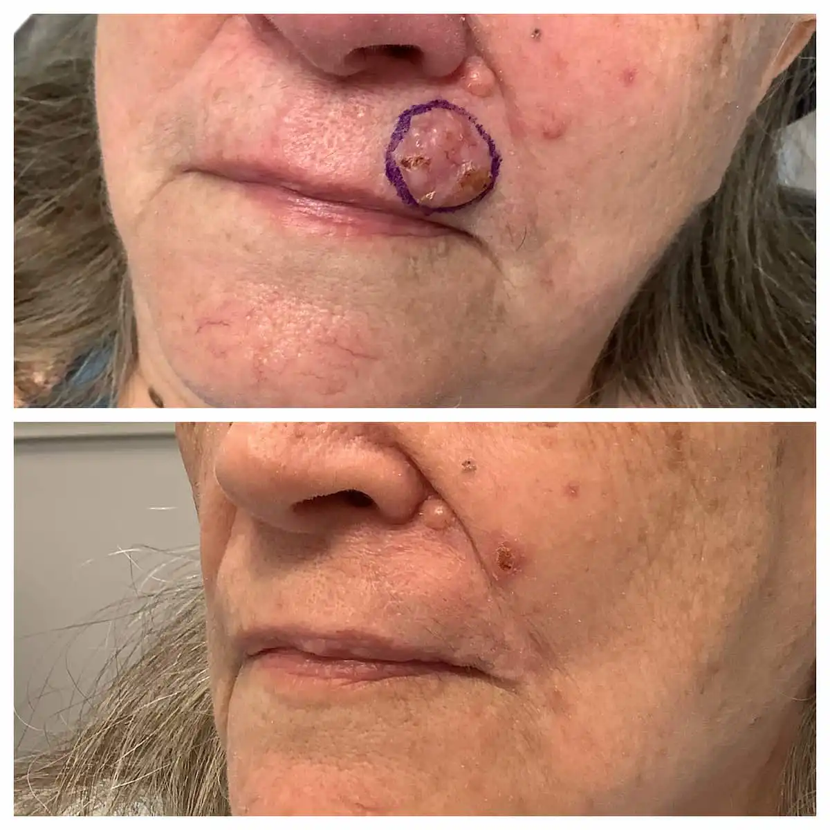
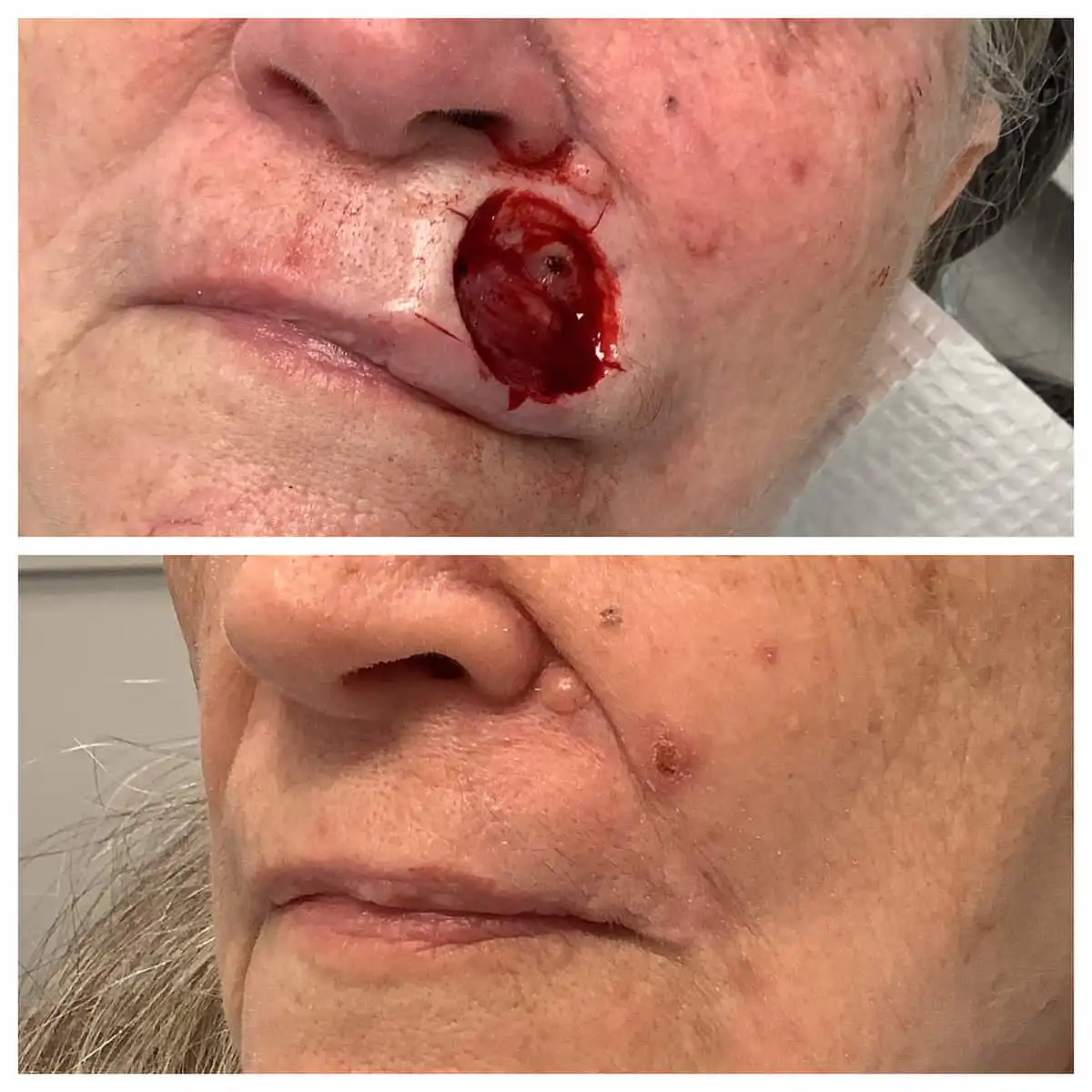
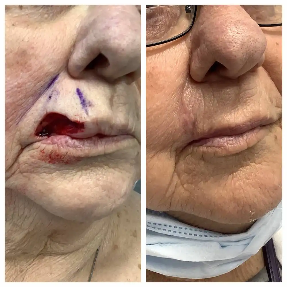
Mohs Surgery & Reconstruction Before and After Photos
Every photo is from a patient of this practice. Skin cancer removed and reconstructed by Dr. Beal.
Common questions
Mohs surgery FAQs.
How long does Mohs surgery take?
Plan to spend several hours with us — sometimes most of the day. Each stage of tissue processing takes roughly 45 minutes to 2 hours, and most of your time is comfortable waiting while your tissue is examined. Many patients bring a book, a snack, or something to watch. The advantage is that you leave the same day knowing the cancer is out and the repair is done.
Does Mohs surgery hurt?
The procedure is performed under local anesthesia, so the area is numb throughout — most patients feel pressure rather than pain. Soreness afterward is normal and typically manageable with simple measures your care team will review with you before you leave.
Will I have a scar?
Any surgery leaves some mark, but Mohs removes the least tissue necessary, and your reconstruction is performed the same day by a surgeon fellowship-trained in facial plastic and reconstructive techniques. The goal is to restore natural contour, color, and texture. Scars typically fade considerably as they mature; results vary by location and skin type.
Is Mohs surgery right for every skin cancer?
No — and that’s by design. Mohs is recommended based on national appropriate-use criteria, typically for skin cancers in high-risk or cosmetically sensitive locations, recurrent tumors, and aggressive subtypes. Some skin cancers are well treated with simpler approaches. Your dermatologist will recommend the option that fits your diagnosis.
Is Mohs surgery covered by insurance?
Mohs surgery is a medically necessary cancer treatment and is typically covered. We accept Medicare and most major private insurance plans. Your exact cost depends on your plan and deductible — call our Troy office at (314) 834-1400 with specific coverage questions.
Diagnosed with skin cancer?
Dr. Beal and our team will walk you through every step — removal, reconstruction, and follow-up, all in one place. Troy, MO — and Chesterfield now open.